Effects Associated with Nanostructure Fabrication Using In Situ Liquid Cell TEM Technology
Corresponding Author: Xin Chen
Nano-Micro Letters,
Vol. 7 No. 4 (2015), Article Number: 385-391
Abstract
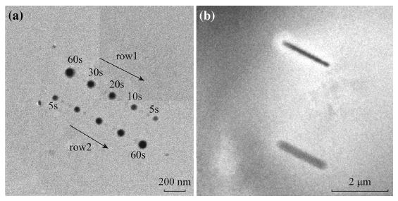
We studied silicon, carbon, and SiC x nanostructures fabricated using liquid-phase electron-beam-induced deposition technology in transmission electron microscopy systems. Nanodots obtained from fixed electron beam irradiation followed a universal size versus beam dose trend, with precursor concentrations from pure SiCl4 to 0 % SiCl4 in CH2Cl2, and electron beam intensity ranges of two orders of magnitude, showing good controllability of the deposition. Secondary electrons contributed to the determination of the lateral sizes of the nanostructures, while the primary beam appeared to have an effect in reducing the vertical growth rate. These results can be used to generate donut-shaped nanostructures. Using a scanning electron beam, line structures with both branched and unbranched morphologies were also obtained. The liquid-phase electron-beam-induced deposition technology is shown to be an effective tool for advanced nanostructured material generation.
Keywords
Download Citation
Endnote/Zotero/Mendeley (RIS)BibTeX
- I. Utke, P. Hoffmann, J. Melngails, Gas-assisted focused electron beam and ion beam processing and fabrication. J. Vac. Sci. Technol., B 26(4), 1197–1276 (2008). doi:10.1116/1.2955728
- R.L. Stewart, Insulating films formed under electron and ion bombardment. Phys. Rev. 45(7), 488–490 (1934). doi:10.1103/PhysRev.45.488
- W.F. van Dorp, C.W. Hagen, A critical literature review of focused electron beam induced deposition. J. Appl. Phys. 104(8), 081301 (2008). doi:10.1063/1.2977587
- J.D. Wnuk, S.G. Rosenberg, J.M. Gorham, W.F. van Dorp, C.W. Hagen, D.H. Fairbrother, Electron beam deposition for nanofabrication: insights from surface science. Surf. Sci. 605(3–4), 257–266 (2011). doi:10.1016/j.susc.2010.10.035
- F. Cicoira, K. Leife, P. Hoffmann, I. Utke, B. Dwir, D. Laub, P.A. Buffat, E. Kapon, P. Doppelt, Electron beam induced deposition of rhodium from the precursor [RhCl(PF3)2]2: morphology, structure and chemical composition. J. Cryst. Growth 265(3–4), 619–626 (2004). doi:10.1016/j.jcrysgro.2004.02.006
- F. Cicoira, P. Hoffmann, C.O.A. Olsson, N. Xanthopoulos, H.J. Mathieu, P. Doppelt, Auger electron spectroscopy analysis of high metal content micro-structures grown by electron beam induced deposition. Appl. Surf. Sci. 242(1–2), 107–113 (2005). doi:10.1016/j.apsusc.2004.08.005
- S.J. Randolph, J.D. Fowlkes, P.D. Rack, Focused, nanoscale electron-beam-induced deposition, etching. Crit. Rev. Solid State Mater. Sci. 31(3), 55–89 (2006). doi:10.1080/10408430600930438
- M.J. Williamson, R.M. Tromp, P.M. Vereecken, R. Hull, F.M. Ross, Dynamic microscopy of nanoscale cluster growth at the solid-liquid interface. Nat. Mater. 2, 532–536 (2003). doi:10.1038/nmat944
- N.D. Jonge, F.M. Ross, Electron microscopy of specimens in liquid. Nat. Nanotechnol. 6, 695–704 (2011). doi:10.1038/nnano.2011.161
- F. Tao, M. Salmeron, In situ studies of chemistry and structure of materials in reactive environments. Science 331(6014), 171–174 (2011). doi:10.1126/science.1197461
- H.G. Liao, K. Niu, H.M. Zheng, Observation of growth of metal nanoparticles. Chem. Commun. 49(100), 11720–11727 (2013). doi:10.1039/c3cc47473a
- X. Chen, C. Li, H.L. Cao, Recent developments of the in situ wet cell technology for transmission electron microscopies. Nanoscale 7(11), 4811–4819 (2015). doi:10.1039/C4NR07209J
- H.M. Zheng, R.K. Smith, Y.W. Jun, C. Kisielowski, U. Dahmen, A.P. Alivisatos, Observation of single colloidal platinum nanocrystal growth trajectories. Science 324(5934), 1309–1312 (2009). doi:10.1126/science.1172104
- E.U. Donev, J.T. Hastings, Electron-beam-induced deposition of platinum from a liquid precursor. Nano Lett. 9(7), 2715–2718 (2009). doi:10.1021/nl9012216
- Y. Liu, X. Chen, W.N. Kyong, J.D. Shen, Electron beam induced deposition of silicon nanostructures from a liquid phase precursor. Nanotechnology 23(38), 385302 (2012). doi:10.1088/0957-4484/23/38/385302
- H.M. Zheng, R.K. Smith, Y.W. Jun, C. Kisielowski, U. Dahmen, P. Alivisatos, Observation of single colloidal platinum nanocrystal growth trajectories. Science 324(5), 1309–1312 (2009). doi:10.1126/science.1172104
- K.W. Noh, Y. Liu, L. Sun, S.J. Dillon, Challenges Associated with in situ TEM in environmental systems: the case of silver in aqueous solutions. Ultramicroscopy 116, 34–38 (2012). doi:10.1016/j.ultramic.2012.03.012
- L.E. Ocola, A.J. Imre, C. Kessel, B. Chen, J. Park, D. Gosztola, R. Divan, Growth characterization of electron-beam-induced silver deposition from liquid precursor. J. Vac. Sci. Technol., B 30, 06FF08 (2012). doi:10.1116/1.4765629
- J.E. Evans, K.L. Jungjohann, N.D. Browning, I. Arsla, Controlled growth of nanoparticles from solution with in situ liquid transmission electron microscopy. Nano Lett. 11(7), 2809–2813 (2011). doi:10.1021/nl201166k
- T.J. Woehl, T.J. Evans, I. Arslan, W.D. Ristenpart, N.D. Browning, Direct in situ determination of the mechanisms controlling nanoparticle nucleation and growth. ACS Nano 6(10), 8599–8610 (2012). doi:10.1021/nn303371y
- G.M. Zhu, Y.Y. Jiang, F. Lin, H. Zhang, C.H. Jin, J. Yuan, D. Yang, Z. Zhang, In situ study of the growth of two-dimensional palladium dendritic nanostructures using liquid-cell electron microscopy. Chem. Comm. 50(67), 9419–9612 (2014). doi:10.1039/c4cc03500c
- J.M. Grogan, N.M. Schneider, F.M. Ross, H.H. Bau, Bubble and pattern formation in liquid induced by an electron beam. Nano Lett. 14(1), 359–364 (2014). doi:10.1021/nl404169a
- X. Chen, L.H. Zhou, P. Wang, H.L. Cao, X.L. Miao, F.F. Wei, A study of electron beam induced deposition and nano device fabrication using liquid cell TEM technology. Chin. J. Chem. 32(5), 399–404 (2014). doi:10.1002/cjoc.201400139
- E.U. Donev, J.T. Hastings, Liquid-precursor electron-beam-induced deposition of Pt nanostructures: dose, proximity, resolution. Nanotechnology 20, 505302 (2009). doi:10.1088/0957-4484/20/50/505302
- P. Abellan, T.J. Woehl, L.R. Parent, N.D. Browning, J.E. Evans, I. Arslan, Factors influencing quantitative liquid (scanning) transmission electron microscopy. Chem. Commun. 50, 4873–4880 (2014). doi:10.1039/c3cc48479c
- X. Chen, K.W. Noh, J.G. Wen, S.J. Dillon, In situ electrochemical wet cell transmission electron microscopy characterization of solid-liquid interactions between Ni and aqueous NiCl2. Acta Mater. 60(1), 192–198 (2012). doi:10.1016/j.actamat.2011.09.047
- N. de Jonge, N.P. Demers, H.D. Demers, D.B. Peckys, D. Drouin, Nanometer- resolution electron microscopy through micrometers-thick water layers. Ultramicroscopy 110(9), 1114–1119 (2010). doi:10.1016/j.ultramic.2010.04.001
- N.M. Schneider, M.M. Norton, B.J. Mendel, J.M. Grogan, F.M. Ross, H.H. Bau, Electron-water interactions and implications for liquid cell electron microscopy. J. Phys. Chem. C 118, 22373–22382 (2014). doi:10.1021/jp507400n
References
I. Utke, P. Hoffmann, J. Melngails, Gas-assisted focused electron beam and ion beam processing and fabrication. J. Vac. Sci. Technol., B 26(4), 1197–1276 (2008). doi:10.1116/1.2955728
R.L. Stewart, Insulating films formed under electron and ion bombardment. Phys. Rev. 45(7), 488–490 (1934). doi:10.1103/PhysRev.45.488
W.F. van Dorp, C.W. Hagen, A critical literature review of focused electron beam induced deposition. J. Appl. Phys. 104(8), 081301 (2008). doi:10.1063/1.2977587
J.D. Wnuk, S.G. Rosenberg, J.M. Gorham, W.F. van Dorp, C.W. Hagen, D.H. Fairbrother, Electron beam deposition for nanofabrication: insights from surface science. Surf. Sci. 605(3–4), 257–266 (2011). doi:10.1016/j.susc.2010.10.035
F. Cicoira, K. Leife, P. Hoffmann, I. Utke, B. Dwir, D. Laub, P.A. Buffat, E. Kapon, P. Doppelt, Electron beam induced deposition of rhodium from the precursor [RhCl(PF3)2]2: morphology, structure and chemical composition. J. Cryst. Growth 265(3–4), 619–626 (2004). doi:10.1016/j.jcrysgro.2004.02.006
F. Cicoira, P. Hoffmann, C.O.A. Olsson, N. Xanthopoulos, H.J. Mathieu, P. Doppelt, Auger electron spectroscopy analysis of high metal content micro-structures grown by electron beam induced deposition. Appl. Surf. Sci. 242(1–2), 107–113 (2005). doi:10.1016/j.apsusc.2004.08.005
S.J. Randolph, J.D. Fowlkes, P.D. Rack, Focused, nanoscale electron-beam-induced deposition, etching. Crit. Rev. Solid State Mater. Sci. 31(3), 55–89 (2006). doi:10.1080/10408430600930438
M.J. Williamson, R.M. Tromp, P.M. Vereecken, R. Hull, F.M. Ross, Dynamic microscopy of nanoscale cluster growth at the solid-liquid interface. Nat. Mater. 2, 532–536 (2003). doi:10.1038/nmat944
N.D. Jonge, F.M. Ross, Electron microscopy of specimens in liquid. Nat. Nanotechnol. 6, 695–704 (2011). doi:10.1038/nnano.2011.161
F. Tao, M. Salmeron, In situ studies of chemistry and structure of materials in reactive environments. Science 331(6014), 171–174 (2011). doi:10.1126/science.1197461
H.G. Liao, K. Niu, H.M. Zheng, Observation of growth of metal nanoparticles. Chem. Commun. 49(100), 11720–11727 (2013). doi:10.1039/c3cc47473a
X. Chen, C. Li, H.L. Cao, Recent developments of the in situ wet cell technology for transmission electron microscopies. Nanoscale 7(11), 4811–4819 (2015). doi:10.1039/C4NR07209J
H.M. Zheng, R.K. Smith, Y.W. Jun, C. Kisielowski, U. Dahmen, A.P. Alivisatos, Observation of single colloidal platinum nanocrystal growth trajectories. Science 324(5934), 1309–1312 (2009). doi:10.1126/science.1172104
E.U. Donev, J.T. Hastings, Electron-beam-induced deposition of platinum from a liquid precursor. Nano Lett. 9(7), 2715–2718 (2009). doi:10.1021/nl9012216
Y. Liu, X. Chen, W.N. Kyong, J.D. Shen, Electron beam induced deposition of silicon nanostructures from a liquid phase precursor. Nanotechnology 23(38), 385302 (2012). doi:10.1088/0957-4484/23/38/385302
H.M. Zheng, R.K. Smith, Y.W. Jun, C. Kisielowski, U. Dahmen, P. Alivisatos, Observation of single colloidal platinum nanocrystal growth trajectories. Science 324(5), 1309–1312 (2009). doi:10.1126/science.1172104
K.W. Noh, Y. Liu, L. Sun, S.J. Dillon, Challenges Associated with in situ TEM in environmental systems: the case of silver in aqueous solutions. Ultramicroscopy 116, 34–38 (2012). doi:10.1016/j.ultramic.2012.03.012
L.E. Ocola, A.J. Imre, C. Kessel, B. Chen, J. Park, D. Gosztola, R. Divan, Growth characterization of electron-beam-induced silver deposition from liquid precursor. J. Vac. Sci. Technol., B 30, 06FF08 (2012). doi:10.1116/1.4765629
J.E. Evans, K.L. Jungjohann, N.D. Browning, I. Arsla, Controlled growth of nanoparticles from solution with in situ liquid transmission electron microscopy. Nano Lett. 11(7), 2809–2813 (2011). doi:10.1021/nl201166k
T.J. Woehl, T.J. Evans, I. Arslan, W.D. Ristenpart, N.D. Browning, Direct in situ determination of the mechanisms controlling nanoparticle nucleation and growth. ACS Nano 6(10), 8599–8610 (2012). doi:10.1021/nn303371y
G.M. Zhu, Y.Y. Jiang, F. Lin, H. Zhang, C.H. Jin, J. Yuan, D. Yang, Z. Zhang, In situ study of the growth of two-dimensional palladium dendritic nanostructures using liquid-cell electron microscopy. Chem. Comm. 50(67), 9419–9612 (2014). doi:10.1039/c4cc03500c
J.M. Grogan, N.M. Schneider, F.M. Ross, H.H. Bau, Bubble and pattern formation in liquid induced by an electron beam. Nano Lett. 14(1), 359–364 (2014). doi:10.1021/nl404169a
X. Chen, L.H. Zhou, P. Wang, H.L. Cao, X.L. Miao, F.F. Wei, A study of electron beam induced deposition and nano device fabrication using liquid cell TEM technology. Chin. J. Chem. 32(5), 399–404 (2014). doi:10.1002/cjoc.201400139
E.U. Donev, J.T. Hastings, Liquid-precursor electron-beam-induced deposition of Pt nanostructures: dose, proximity, resolution. Nanotechnology 20, 505302 (2009). doi:10.1088/0957-4484/20/50/505302
P. Abellan, T.J. Woehl, L.R. Parent, N.D. Browning, J.E. Evans, I. Arslan, Factors influencing quantitative liquid (scanning) transmission electron microscopy. Chem. Commun. 50, 4873–4880 (2014). doi:10.1039/c3cc48479c
X. Chen, K.W. Noh, J.G. Wen, S.J. Dillon, In situ electrochemical wet cell transmission electron microscopy characterization of solid-liquid interactions between Ni and aqueous NiCl2. Acta Mater. 60(1), 192–198 (2012). doi:10.1016/j.actamat.2011.09.047
N. de Jonge, N.P. Demers, H.D. Demers, D.B. Peckys, D. Drouin, Nanometer- resolution electron microscopy through micrometers-thick water layers. Ultramicroscopy 110(9), 1114–1119 (2010). doi:10.1016/j.ultramic.2010.04.001
N.M. Schneider, M.M. Norton, B.J. Mendel, J.M. Grogan, F.M. Ross, H.H. Bau, Electron-water interactions and implications for liquid cell electron microscopy. J. Phys. Chem. C 118, 22373–22382 (2014). doi:10.1021/jp507400n

